Female Reproductive System Of Cow
Anatomy And Physiology Of Reproductive System :
Female reproductive tract of various farm animals are similar to the cow but differ primarily in the shape of uterus and cervix.
Female tract consists of :
➤Vulva
➤Vagina
➤ Cervix
➤Uterus
➤Uterine Horns
➤ Fallopian Tube
➤Ovaries

Vulva

- Exterior portion and opening of the female reproductive tract.
Clitoris
- This organ is responsible for stimulating female orgasm. The clitoris is the sensory and erectile organ of the female(sensory organ analogous to the male's penis). It is located just inside the vulva. Stimulation of the clitoris causes contractions that move semen into the female reproductive tract.
Vagina
- The vagina serves as the female organ of copulation (receptacle for the male's penis) at mating and as the birth canal at parturition. It is the passage but the cervix and vulva. The lining is moist in estrous and dry when the animal is not in estrous.
- It is the opening of the bladder and serves as a urinary tract.
- Semen is deposited in this region in cows ana whereas mares and sows have semen deposited in the cervix.
- During mating, millions of sperms are deposited in the female vagina.
- Sperms travel from the vagina through the cervix and uterus and fallopian tube.
- Cow 35-42 cm
- Ewe 12-17 cm
- Sow 16-23 cm
- Mare 30-47 cm
Cervix:
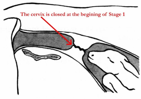
The cervix is the lower outlet of the uterus. It is composed primarily of the connective tissues that contribute to the gateway between the uterus and the vagina. Like the rest of the reproductive tract, the cervix is lined with mucosal cells. These cells make significant changes as the animal goes from one estrous cycle to another and during gestation or pregnancy.
The cervix has annular rings which are 4-5 in cows. Cervix ix composed of thick connective tissue and has thick walls that allow a passageway for sperms at mating and expulsion of the fetus at the time of birth. During pregnancy, the cervix is filled with a thick mucous secretion known as a cervical plug which protects the uterus from infections entering the vagina.
Cervix connects and divides the uterus and vagina.
Cervix is the mouth of the womb:
- The opening into the uterus which sperm must pass to fertilize the egg.
Cervix stretches during birth to allow passage of newborn.
Uterus:

- Site of embryonic development during gestation and provide nourishment for the fetus.
- The uterus of cow is bipartile, while the uterine horns are relatively long and well developed.
- The uterus undergoes tremendous changes in size, structure, and position during pregnancy. After parturition, uterine involution occurs and the uterus undergoes to its normal size.
The Uterine wall consists of:
- Serous membrane
- Myometrium-internal circular muscle-external longitudinal muscle-connective tissue
Uterine Horns:
- Two branches of the uterus
- Fallopian tubes or oviducts are located at the end of each horn.
- Lined with microscopic cilia-help guide cells to the horn
- Usually, the size of sperm and ovum unite.
Fallopian Tube:(Oviduct)

Sperms and egg unite in the fallopian tube. Transport embryos to the uterus. The oviduct can be divided into three parts namely the Infundibulum, Ampulla, and Isthmus.
- The oviduct or the fallopian tubes are two tubes that carry the ova from the ovaries to the uterus. The oviducts are close, but not attached to the ovaries.
- The funnel-shaped end of each oviduct that is close to the ovary is called the infundibulum. At ovulation, the follicles rupture, releasing an ovum that is caught by the infundibulum.
- After copulation, sperm move through the uterus to the oviduct. Fertilization of the ovum occurs in the upper end of the oviduct. The Zygote or the Fertilized egg moves to the uterus about 2-4 days after fertilization.
Ovaries:
A female mammal will typically have two ovaries that produce female gametes (ova or egg). A gamete is a sex cell that can unite with other sex cells. Egg contains 1/2 of the genetic makeup.
- The ovaries are stimulated by a hormone called the follicle-stimulating hormone (FSH) and produce a follicle where the egg grows and matures.
- Within each ovary, there are hundreds of tiny follicles or cavities. The ova are produced in the follicles, the largest single cell in the body. When the egg matures, the follicle ruptures and releases the egg-Ovulation
- The ovaries also produce the female sex hormones Estrogen & Progesterone.
Comments
Post a Comment